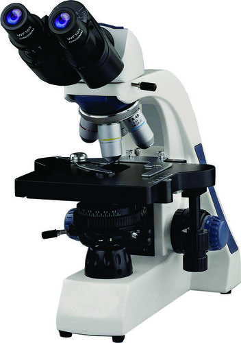
Computer Assisted Semen Analyzer
Product Details:
- Product Type Computer Assisted Semen Analyzer
- Color White
- Usage Laboratory
- Theory
- Drawtube Binocular
- Click to View more
Computer Assisted Semen Analyzer Price And Quantity
- 10000.00 - 50000.00 INR/Piece
- 10000 INR/Piece
- 150 Piece
Computer Assisted Semen Analyzer Product Specifications
- Computer Assisted Semen Analyzer
- Laboratory
- White
- Binocular
Computer Assisted Semen Analyzer Trade Information
- 10 Piece Per Month
- 10-30 Days
- Contact us for information regarding our sample policy
- Australia South America Eastern Europe Western Europe Middle East Central America Asia North America Africa
- All India
Product Description
Specifications:-
* Head: Binocular Observation head, inclined at 30 degree, rotatable at 360 degree, interpupillary distance from 55 to 75, +/- 5 diopter adjustable.
* Eyepieces: Plan 10x/18mm (FOV, pair) High Eyepoint wide field Eyepieces.
* Objectives: Semi Plan Achromatic Objective 4x, N.A. 0.10/ W.D. 18.5mm,
Semi Plan Achromatic Objective 10x, N.A. 0.25/ W.D. 10.6mm,
Semi Plan Achromatic Objective 40x, N.A. 0.65 (spring)/ W.D. 0.6mm,
Semi Plan Achromatic Objective 100x, N.A. 1.25 (Oil, spring)/ W.D. 18.5mm.
* Nosepiece: Revolving Quardruple Nosepiece on Ball Bearing.
* Focusing System: Coaxial Coarse and fine focusing system. Re focusing lever & tension adjustment ring. The slow motion has 1 division 0.002mm.
* Mechanical Stage: Double Plate mechanical stage size 140mm x 155mm and having low positioned coaxial coarse in ball bearing. Coarse movement 55mm x 75mm.
* Sub Stage Condenser: Abbe condenser N.A. 1.25 sawing out filter holder. Iris diaphragm.
* Illumination System: Built in base illumination system with 3W LED/Halogen bulb and adjustable brightness.
Camera:-
Microscopy HDMI Digital Color Camera 3MP with core Processor for Screen Microscope Image display system with driver software 64 bit, C mount optical with 0.5x relay lens. HDMI USB 3.0 SD card connection cable. Basic High 1K resolution. PC connectivity USB 3.0 interface, 100 micro sec to 30 sec increment exposure control, 30 fps live frame rate.
Laptop:-
Dell/HP/Lenovo/Acer/Samsung/LG and other branded Laptop 14 inch IPS LED backlit Touch display with Intel Core i5 processor (3.0), 8 GB RAM, 64 GB of SSD Storage, Wifi, bluetooth, Ethernet, Graphic HD, USB 3.0 slot, 6 USB port, HDD, DVD writer, window 10 professional, HDMI connector, VGA, External I/O 6 USB ports.
Printer:-
Monochrome laser and color jet desk printer, Max class of 4A (210x297mm)/Legal (216x356mm), min custom (76x127mm) Media size. Print Speed up to 75 ppm, 1200 x 1200 dpi max resolution, Max 800 sheets capacity.
Sperm Analysis Software:-
Introduction of Sperm Analysis:
Sperm Analysis is simple image analysis software for microscopy with an intuitive user interface and simple to use navigation with a suite of image processing techniques, measurements and enhancements tool that set it apart from other mainstream software.
Such Image processing software are how being extensively used in a number of diverse fields such a medicine, biological research, cancer research, drug testing etc.
Motility:
* Motility module perform motility (motion) analysis of sperm from the input video of any time period.
* Motility analysis is determined by computing the sperm cells movement and its path in the frame.
* Motility measures the count and percentage of fast moving, slow moving and dead (immotile) sperm.
* Also measure the overall count and percentage of motile and immotile sperm.
Morphology:
* Morphology analysis perform parametric study of sperm.
* The module perform following measurements:-
Area of sperm Head
Perimeter of sperm Head
Its similarity ratio with circle and ellipse.
Acrosome content percentage in the sperm head.
Small and big axis (major and minor axis) of sperm head.
* Identification of type defect as per WHO standard:-
Head defect, Tail defect, Neck and mid-piece defect Cytoplasmic droplet
DNA Fragmentation:
* Perfect DNA Fragmentation analysis of sperms based on input image.
* Identification of Sperm in Types:-
Normal Sperm, Fragmented Sperm Degraded Sperm
* Computer count and percentage of normal, fragmented and degraded sperm
Vitality:
* Vitality analysis perform identification of live or dead sperms.
* Measures count and percentage of live and dead sperms.
Report:
Patient Name:______________________
Evaluation date:____________________
Report No:________________________
Motality Test:-
Total Sperm Analyzed count : 335
Rapid Motality Sperm Count : 123
Slow and Sluggish Motality Count : 100
Immotile Sperm count : 112
Motalite Sperm : 223
Rapid Motality Percentage : 36.7164
Slow and Sluggish Motality Percentage : 29.8507
Immotile Sperm Percentage : 33.4528
Motile Sperm Percentage : 66.5672
Morphology Test:-
Area of Head : 2231.5
Perimeter of Head : 185.439
Big Axis : 67.4689
Small Axis : 43.267
Form factor circle : 0.623918
Form factor Ellipse : 0.987396
Acrosome Level : 0.991261
Type Classifier :
DNA Fragmentation Test:-
Total Sperm Analyzed : 14
Normal Count : 7
Fragmented Count : 69
Degraded Count : 1
Normal Percentage : 50
Fragmented Percentage : 42.8571
Degrade Percentage : 7.14286
Vitality Test:-
Total Sperm Analyzed : 46
Live count : 21
Dead count : 25
Live Percentage : 45.6522
Dead Percentage : 54.3476
Remark:______________ Approved By:______________________
Annotation:-
Annotation features allow quick and easy documentation of your image tool. The Available Tools are Line between 2 Points, Horizontal line, Vertical Line, Free hand line, Two parallel line, Perpendicular Line, Square, Circle, Text, Arrow, Grid, Cross Lines from center, scale bar, highlighter. The Text color and thickness of line can be designated
Measurement:-
A large number of measurement tools are available to perform on stored images on no live display. Cross line or grid of 10x10 can be deployed there on live display or on capture images. Line measurement tools are: Length of line, distance between two points, Length of the vertical lines, Length of the Horizontal lines, Freehand tool to measure length of an irregular object, area of and perimeter of irregular shapes, perpendicular line measures the distance from any baseline/fiducial line, distance between two parallel lines, measures the diameter of circle created by points, area and perimeters of irrigular shapes, Arc, ellipse, circle measurements including definition of center point, Chord length, Sweep angle & radius.
Manual Stitcher:-
The software is designed to stitch overlapped image tiles by moving stage precisely step by step to cover the entire specimen and sub segments of stitching of the data blocks into one image. It has an automatic algorithm for homogeneous illuminations using shading (black back ground) correction. Stitching options are available for few images as well as whole samples with resolution up to several giga-pixel.
Ideal for large tissue sample, this ensure reproducblity while taking the guesswork out of tilting experiments.
Montage (Z-Staking):-
This module is a digital image processing method, which combine multiple image taken at different focal distances (Z-staking) to provide a composite image with a greater depth of field (i.e. thickness of the plane of the focus) than any of the individual source images. It is particularly useful for capturing in focus image of objects under light magnifications. With this method you can extract specific parts of the image info three dimensional images.
Time Lapse:-
Time Lapse Acquisition investigates change in specimens or materials over time by acquiring images at predefined intervals. It supports tiff, bmp & JPG file formats.
It also includes an auto save feature by DD/MM/YY/hour/minute/second play your time lapse images as a movie to view the movement and other activities.
Overlay:-
Overlay this application, Image overlay module, acquires, enhances and documents multiple wavelength, fluorescence microscopy images.

