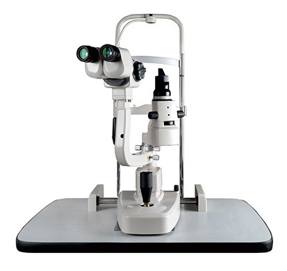
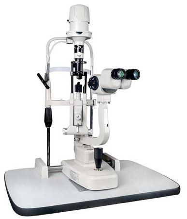
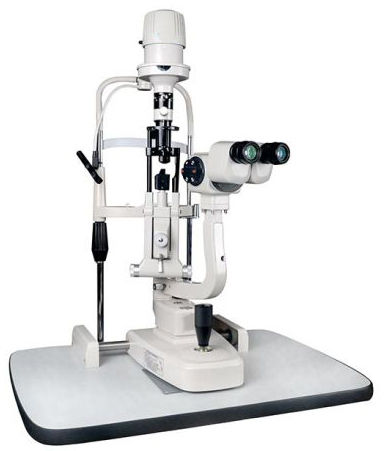
Slit Lamp 5 Step
Product Details:
- Product Type Slit Lamp 5 Step
- Usage Laboratory
- Color White
- Theory Other
- Drawtube Binocular
- Click to View more
Slit Lamp 5 Step Price And Quantity
- 10000.00 - 50000.00 INR/Piece
- 150 Piece
- 10000 INR/Piece
Slit Lamp 5 Step Product Specifications
- Slit Lamp 5 Step
- White
- Other
- Laboratory
- Binocular
Slit Lamp 5 Step Trade Information
- 10 Piece Per Month
- 10-30 Days
- Contact us for information regarding our sample policy
- Australia North America South America Eastern Europe Western Europe Middle East Central America Asia Africa
- All India
Product Description
Slit Lamp
The Slit Lamp is essentially a simple and generally under- user piece of equipment. It consists of an illumination system and a binocular observation system, which when correctly aligned with result in a coincidental focus of the slit and microscope.
Illumination System
Basically a short focus projector projecting an image of the illuminated slit aperture on to the eye. This part of the system should be flexible to allow various sizes and shape of slit beam. Usually a rheostat is incorporated and the lamp house can be rotated and the lamp house can be rotated. Neutral density, cobalt blue and red free filters are usually available, and occasionally a diffuses and Polarizes.
Using the Slit Lamp
Commence the examination using the 10x eyepieces and the lower powered objectives i.e. 1x. Use the lowest voltage setting on the transformer. Select the longest slit length by means of the appropriate lever. Adjust the chin rest by means of control so that the patient's eyes are approximately level with the black marker on the side of the head rest. Adjust the height of the slit lamp until the slit beam is centered vertically on the patients eyes. Focus the slit beam in the eye by moving the joystick either towards or away from patients. Coarse positioning can be effected without using the microscope but critical focusing should be carried out whilst viewing through the microscope. The slit width is variety by rotating either the left hand or right hand knurled control. To vary the angle between illumination and microscope use one or other of these same controls are handles.
The slit should be set primarily in the vertical position, but any desired inclination can be achieved by mean of the ball handle (notches at 45 degree, 90 degree, 135 degree, stop at 0 to 180 degree). By tripping the latch and tilting the slit lamp column, the beam can be introduced from as much as 20 degree below the horizontal. This is mainly used for carrying out goniscopy.
For observation by sclerotic scatter or other dissociated forms of examination the centering screw is loosened, so that the slit image can be moved away from the center of the field of observation. The Image is centered again by tightening the screw. Carry out the techniques described below.
Specifications
* Viewing ocular Binocular, Galilean type
* Magnifications Change 5 Steps Drum Rotation type Magnification
* Eyepieces Wide Field 12.5x
* Objectives 0.4x, 0.6x, 1x, 1.6x, 2.5x
* Total Magnifications 5x, 7.5x, 12.5x, 20x, 30x
Illumination unit
* Slit Image Rotation 0 to 180 degree
* Tilting Illumination 5 degree to 20 degree
* Filter Disc. Cobalt Filter, Red free, Yellow Filter natural Density and open aperture
Diaphragm for infinitely variable slit lengths
* Slit Diaphragm Disc. Slit Aperture of 12, 9, 7,3, and 0.2 mm and a wedge shaped Diaphragm
of Infinitely variable slit lengths
* Halogen Lamp 12 Volts 50 Watts
Standard Accessories
* Replacing Mirror Plastic Breath Shield
* Testing Rod Spare Lamp

Price:
- 50
- 100
- 200
- 250
- 500
- 1000+


